Abstract
The cross-sectional imaging appearance of the abdominal vasculature during acquisition with Valsalva maneuvers can have unfamiliar appearances mimicking pathologic processes resulting in incorrect diagnosis risking unnecessary additional imaging and follow-ups. Herein, we report a case of unique arterial findings generated by the Valsalva maneuver during computed tomography acquisition in a patient undergoing evaluation for inguinal hernia.
Keywords
Aorta; Intra-abdominal pressures; Valsalva
Article text
Case Presentation
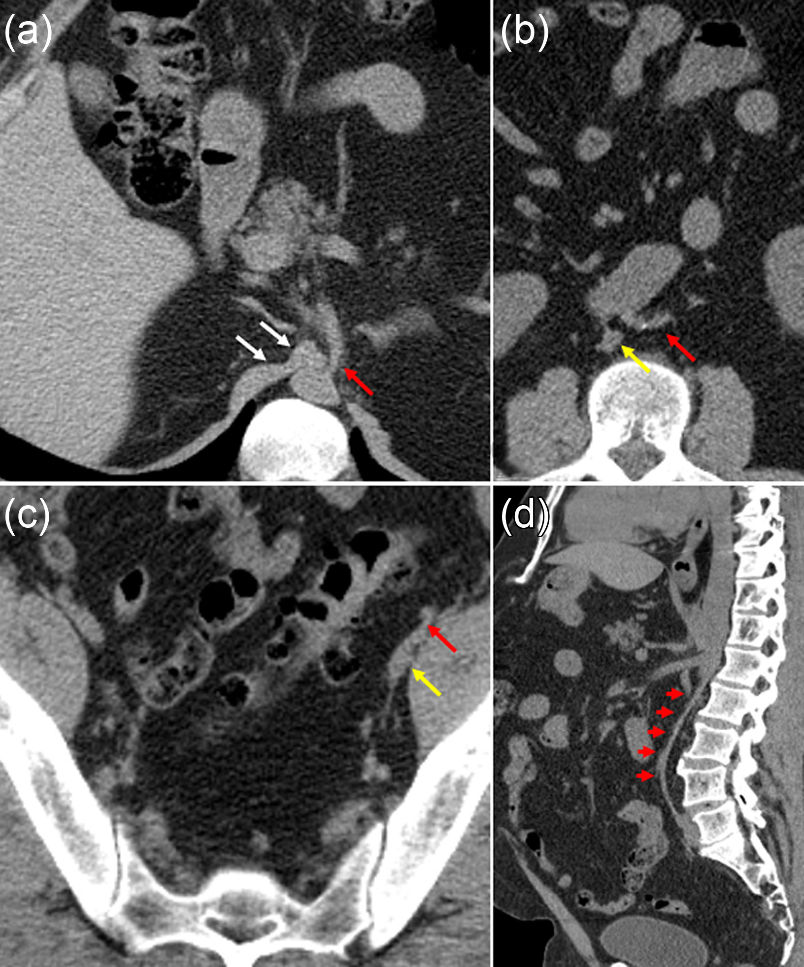
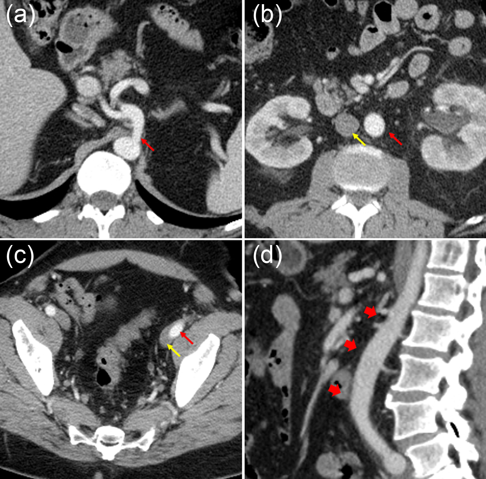
A 59-year-old male with a body mass index (BMI) of 32.7 kg/m2 presented to his primary care physician with a 2-week history of swelling and discomfort in the left groin. An inguinal hernia was not evident on the physical examination, and therefore, an unenhanced computed tomography (CT) of the abdomen and pelvis was performed on an outpatient basis. A CT scan showed a small fat-containing left inguinal hernia as well as an incidental finding of marked narrowing of the abdominal aorta and the iliac arteries (Figure 1). This was interpreted as aortic narrowing of unclear etiology with retroperitoneal lipomatosis as a potential differential. The patient then presented 3 months later to the emergency department with complaints of fever, dysuria, and vomiting. Urinalysis was consistent with urinary tract infection. A contrast-enhanced CT of the abdomen and pelvis was performed to evaluate for nephroureterolithiasis and pyelonephritis. The CT demonstrated an obstructing left ureteral stone with hydronephrosis. In contrast to the CT obtained 3 months earlier, the abdominal aorta and the iliac arteries were normal in caliber (Figure 2).
Discussion
A commonly used imaging protocol for patients undergoing CT for the detection of abdominal hernias is to perform the scan during the Valsalva maneuver. This has shown to be sufficient for the detection of hernias in up to 100% of cases.1 Valsalva maneuvers such as coughing, vomiting, retching, or sneezing result in a transient increase in the intra-abdominal pressure (IAP), with rapid normalization of pressure when the maneuver is ceased.2
Intra-abdominal pressures above 12 mm Hg, measured by intra-vesicular manometry, are defined as intra-abdominal hypertension (IAH). The common imaging finding associated with IAH is the elevation of the left hemidiaphragm. Abdominal compartment syndrome (ACS) is a surgical emergency with a high mortality rate and is defined as an IAP of ≥20 mm Hg with evidence of organ failure. The common imaging findings associated with ACS are compression of major abdominal venous structures including the inferior vena cava (IVC), renal veins, and iliac veins. Major arterial compression has not been described in ACS as the pressure needed to compress the major arteries is very high and has to exceed at least the diastolic pressure.3 Cobb et al4 measured IAPs in 20 healthy young adults using intra-vesicular manometry and reported an increase in the IAPs up to 107 mm Hg with coughing. In addition, patients with a higher BMI were noted to generate significant elevation in IAP.4 Similarly, Iqbal et al2 reported an increase in the IAP of up to 290 mm Hg with vomiting. In our patient, the blood pressure was 130/80 mm Hg prior to his CT scan, which suggests that the IAP must have been greater than 80 mm Hg (well above the ACS range) to have compression of the major arteries.
Valsalva maneuvers result in massively increased intra-abdominal pressures above the patient’s diastolic pressure resulting in marked narrowing of aortoiliac arteries and compression of the IVC and renal veins. Clinical and imaging features that help differentiate this benign physiologic entity from pathologic aortoiliac narrowing include transient nature, absence of symptoms, absence of aortic wall thickening and periaortic inflammatory changes, and absence of mass. Awareness of this entity and its imaging findings should help radiologists in making a confident diagnosis to avoid unnecessary follow-ups and imaging.
Figures
Declarations
- Informed Consent
- Verbal informed consent was obtained from the patient who agreed to take part in the study.
- Peer Review
- Externally peer-reviewed.
- Author Contributions
- Concept - I.M., H.K.; Design - I.M., H.K.; Supervision - I.M.; Resources - I.M., H.K.; Materials - I.M., H.K.; Data Collection and/or Processing - I.M.; Analysis and/or Interpretation - I.M., H.K.; Literature Search - I.M., H.K.; Writing Manuscript - I.M.; Critical Review - I.M.
- Declaration of Interest
- The authors have no conflict of interest to declare.
- Funding
- The authors declared that this study has received no financial support.
References
- Jaffe TA, O'Connell MJ, Harris JP, Paulson EK, Delong DM. MDCT of abdominal wall hernias: is there a role for valsalva’s maneuver? AJR Am J Roentgenol. 2005;184(3):847-851. [CrossRef]
- Iqbal A, Haider M, Stadlhuber RJ, Karu A, Corkill S, Filipi CJ. A study of intragastric and intravesicular pressure changes during rest, coughing, weight lifting, retching, and vomiting. Surg Endosc. 2008;22(12):2571-2575. [CrossRef]
- Krol JJ, Hallett RL. Transient, marked flattening of the abdominal aorta, iliac arteries, and venous structures during active vomiting: report of a case demonstrated by MDCT. Abdom Imaging. 2011;36(5):582-585. [CrossRef]
- Cobb WS, Burns JM, Kercher KW, Matthews BD, Norton HJ, Heniford BT. Normal intraabdominal pressure in healthy adults. J Surg Res. 2005;129(2):231-235. [CrossRef]
Cite this article
Irfan Masood, Hasan Khan. Vanishing Aorta: A Case of Transient Marked Flatting of the Abdominal Aorta and Iliac Arteries. Imaging and Intervention. 2021;1(3):74–75. doi:10.5152/iai.2022.22001